Digital dentistry clinic · central Niš
We measure, we plan,then we treat
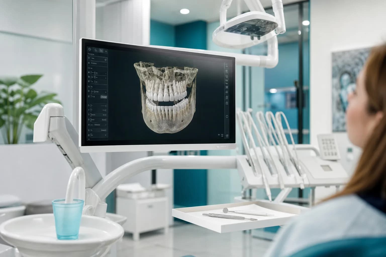
At Oris Clinic every decision starts with data: a CBCT scan, a digital impression and an examination under magnification. We show you the plan on screen, and you receive the images and report too.
- CBCT and digital scanning on site
- Implants planned around the future crown
- A copy of your images and plan
Clinic
How we work
CBCT on site
We take three-dimensional scans on site, with no referral to another facility.
Written plan and costs
Before treatment you receive phases, alternatives and estimated costs on paper or as a PDF.
Microscope in endodontics
We treat root canals under magnification, with images we can show you.
Room for a second opinion
With a copy of your images you are free to seek another dentist's opinion as well.
Treatments
Treatment that starts with measuring
From 3D diagnostics and guided implants to CAD/CAM crowns and microscope root canal treatment: every area uses the same digital records and the same order of work.
| Treatment | Price | ||
|---|---|---|---|
| Guided dental implant surgeryAn implant whose position, depth and angle are set on a CBCT scan and a digital model, then transferred to the mouth with a surgical guide where indicated.from RSD 85,000First visit: 60 min | An implant whose position, depth and angle are set on a CBCT scan and a digital model, then transferred to the mouth with a surgical guide where indicated. | from RSD 85,000 | First visit: 60 min |
| CAD/CAM crowns and prosthodonticsCrowns, inlays, onlays, bridges and dentures made from a digital impression; single ceramic restorations, where indicated, even within the same visit.from RSD 42,000First visit: 60 min | Crowns, inlays, onlays, bridges and dentures made from a digital impression; single ceramic restorations, where indicated, even within the same visit. | from RSD 42,000 | First visit: 60 min |
| Urgent dental care with prompt diagnosticsA same-day examination whenever the schedule allows, with imaging that shows the cause of the pain, and treatment that deals with the acute problem.from RSD 3,500First visit: 30 min | A same-day examination whenever the schedule allows, with imaging that shows the cause of the pain, and treatment that deals with the acute problem. | from RSD 3,500 | First visit: 30 min |
| Check-up and 3D diagnosticsA thorough examination with a digital scan, radiographs as indicated and CBCT where needed, after which you receive a report and plan with a copy of all records.from RSD 4,000First visit: 60 min | A thorough examination with a digital scan, radiographs as indicated and CBCT where needed, after which you receive a report and plan with a copy of all records. | from RSD 4,000 | First visit: 60 min |
Clinic and equipment
The clinic and its equipment
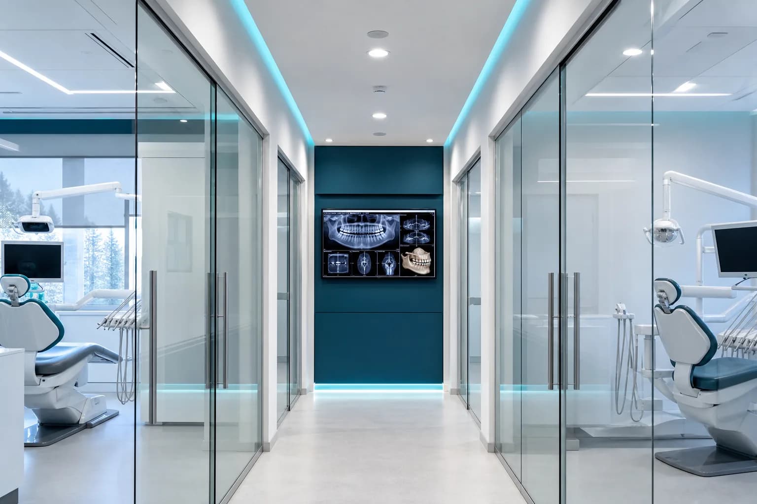
The equipment at Oris Clinic was chosen with one question in mind: does it help us measure, plan and carry out treatment more precisely?
- Intraoral scannerThe scanner records the teeth and gums with a small camera and creates an accurate three-dimensional model within minutes. That model is the starting point for planning implants, CAD/CAM crowns, veneers and aligners, and it is stored in your digital record for comparison at check-ups.
- Digital intraoral X-rayA sensor placed in the mouth captures an image of one to three teeth, which appears on screen immediately. We use it to check for decay between teeth, follow root canal treatment and check crown margins, with a low radiation dose adjusted to the patient's size.
- CBCT unit with adjustable field of viewCBCT produces a three-dimensional image of the jaw while the patient stands or sits still for a few tens of seconds. We choose the field of view to fit the question, from one tooth to part of a jaw or both jaws, so the dose stays as low as possible while the image is sufficient for implant planning, endodontics or clarifying an unclear finding.
Our team
Dentists working from the same data
Our surgeon, prosthodontist, endodontist and children's dentist work from shared digital records, so nothing gets lost between treatment rooms.
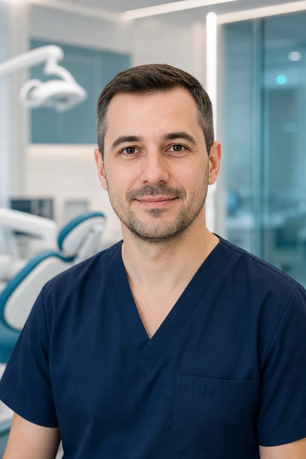
Specialist in oral surgery
Dr Dušan Mitić
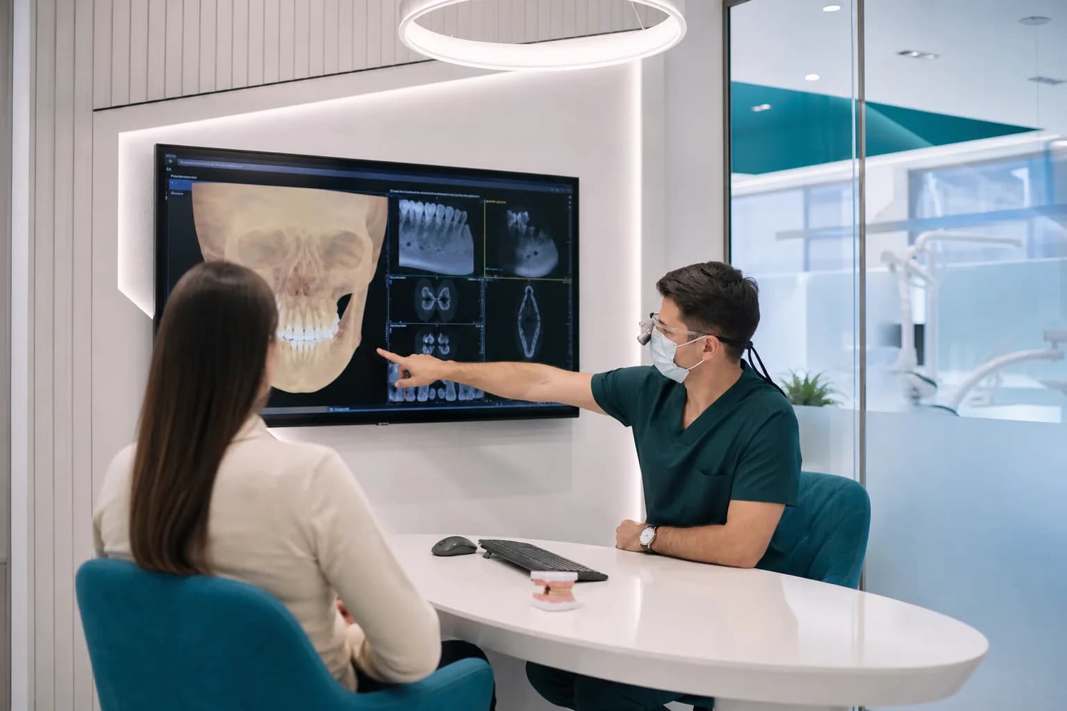
Dr Dušan Mitić plans every implant on a CBCT scan and a digital model before scheduling surgery. He shows patients the plan on screen, slice by slice.
Book with this dentist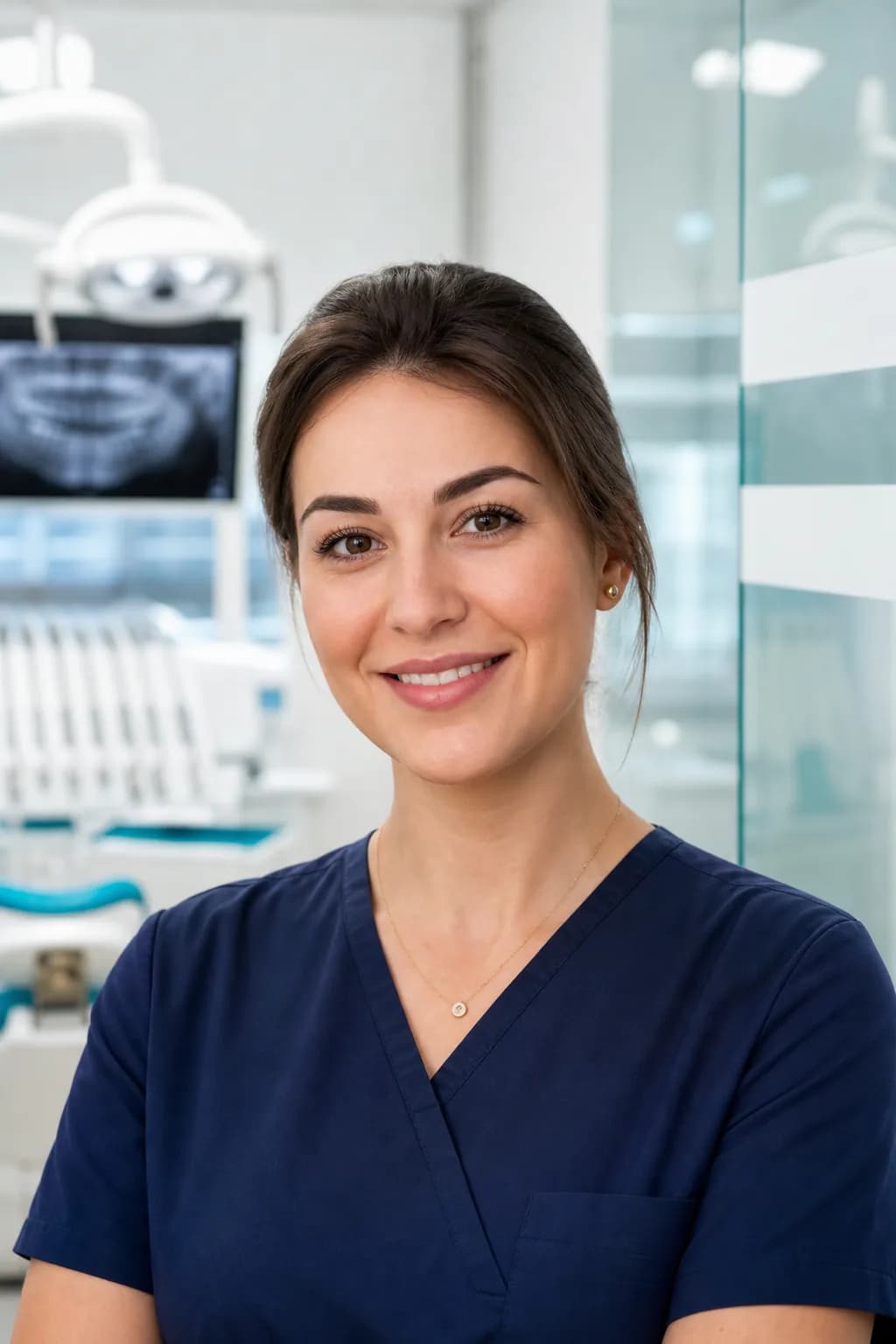
Dr Tamara Živković
Specialist in prosthodontics
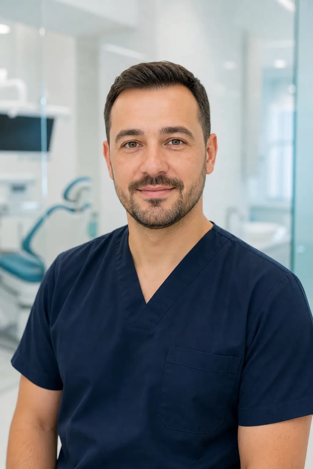
Dr Vladimir Cvetković
Specialist in restorative dentistry and endodontics
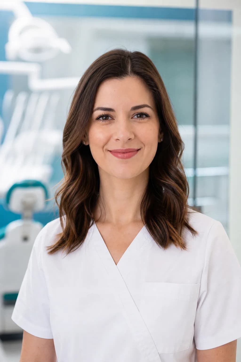
Dr Ivana Stanković
Specialist in paediatric and preventive dentistry
Patient experiences
How patients describe treatment with the scan on screen
Dr Mitić showed me on screen how much bone I have and where the nerve runs, and for the first time I understood why I had been told an implant would not be straightforward. The placement went more calmly than I expected, and I took a copy of the scan and plan home with me.
I came in one morning with a broken molar and left in the afternoon with a ceramic crown. Between the scan and the fitting I had a coffee in the waiting room and watched the crown being milled. The tooth was a little sensitive to cold for the first couple of days, and after that nothing.
My five-year-old son was afraid of dentists after one bad experience. Dr Stanković let him hold the camera himself and look at his teeth on screen, and in the end he opened his mouth without any coaxing. We were also given a short written guide to brushing.
When you need help today
During opening hours we see urgent patients on the same day whenever the schedule allows. Call us before you come so we can assess the urgency and get imaging ready.
FAQ
Answers to the questions we hear most
Here you will find general information about treatment, imaging, prices and travelling from abroad. Assessing your own case requires an examination and, if needed, imaging.
Most procedures are carried out under local anaesthetic, so patients usually feel pressure and vibration during treatment, but not pain. Before we begin, we show you on screen what we are going to do and agree a signal for a pause. Some mild tenderness is possible once the anaesthetic wears off, depending on the procedure and the clinical findings.
For single ceramic crowns, inlays and onlays it is often possible: we scan the tooth, design the restoration and mill it in the clinic during one visit of two to three hours. When the tooth margin lies below the gum, when root canal treatment is needed or when several connected teeth are involved, working in stages is the better choice. Whether one-visit treatment is indicated is decided after the examination and radiograph.
A panoramic radiograph shows the jaw in two dimensions, so it does not reliably show the width of the bone or the exact distance to the nerve and sinus. CBCT provides a three-dimensional image on which we measure the bone in every direction and plan the implant position before surgery. We choose the field of view for the area being planned, to keep the dose as low as possible.
You can book through the form on the website, by phone or by message, weekdays from 8 am to 8 pm and Saturdays from 8 am to 2 pm. After your request we send a confirmation with the time and address, and a reminder the day before. If you have earlier X-rays or scans, bring them or send them in advance.
Booking
Book an examination
Choose the type of visit and a time that suits you. We are open weekdays from 8 am to 8 pm and Saturdays from 8 am to 2 pm.